Renal colic assessment Safer Care Victoria
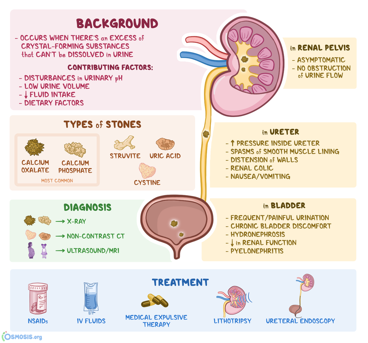
Urinary stones are the main cause of renal colic. If the stone restricts your flow of urine, it can cause increased pressure and inflammation. Stones can form in your urinary tract for several reasons, including: Dehydration. Excess calcium in your pee. Excess protein in your diet. Certain medications.

Figure 1 from The Diagnosis and Management of Patients with Renal Colic across a Sample of US
Unspecified renal colic. ( N23) N23 is a billable diagnosis code used to specify a medical diagnosis of unspecified renal colic. The code is valid during the current fiscal year for the submission of HIPAA-covered transactions from October 01, 2023 through September 30, 2024. Unspecified diagnosis codes like N23 are acceptable when clinical.
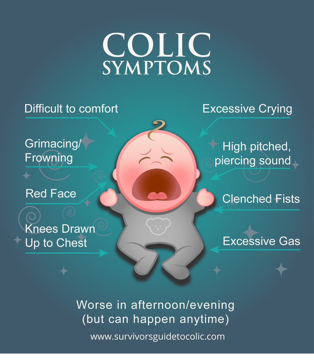
About Colic — Survivor's Guide to Colic
To assess whether ultrasonography (US) with or without plain abdominal radiography (kidney, ureter, bladder [KUB] radiography) can replace intravenous urography (IVU) in detection of acute urinary tract obstruction, 101 consecutive patients with renal colic were evaluated with US followed immediately by IVU. Receiver operating characteristic (ROC) curves for US diagnosis of acute urinary tract.
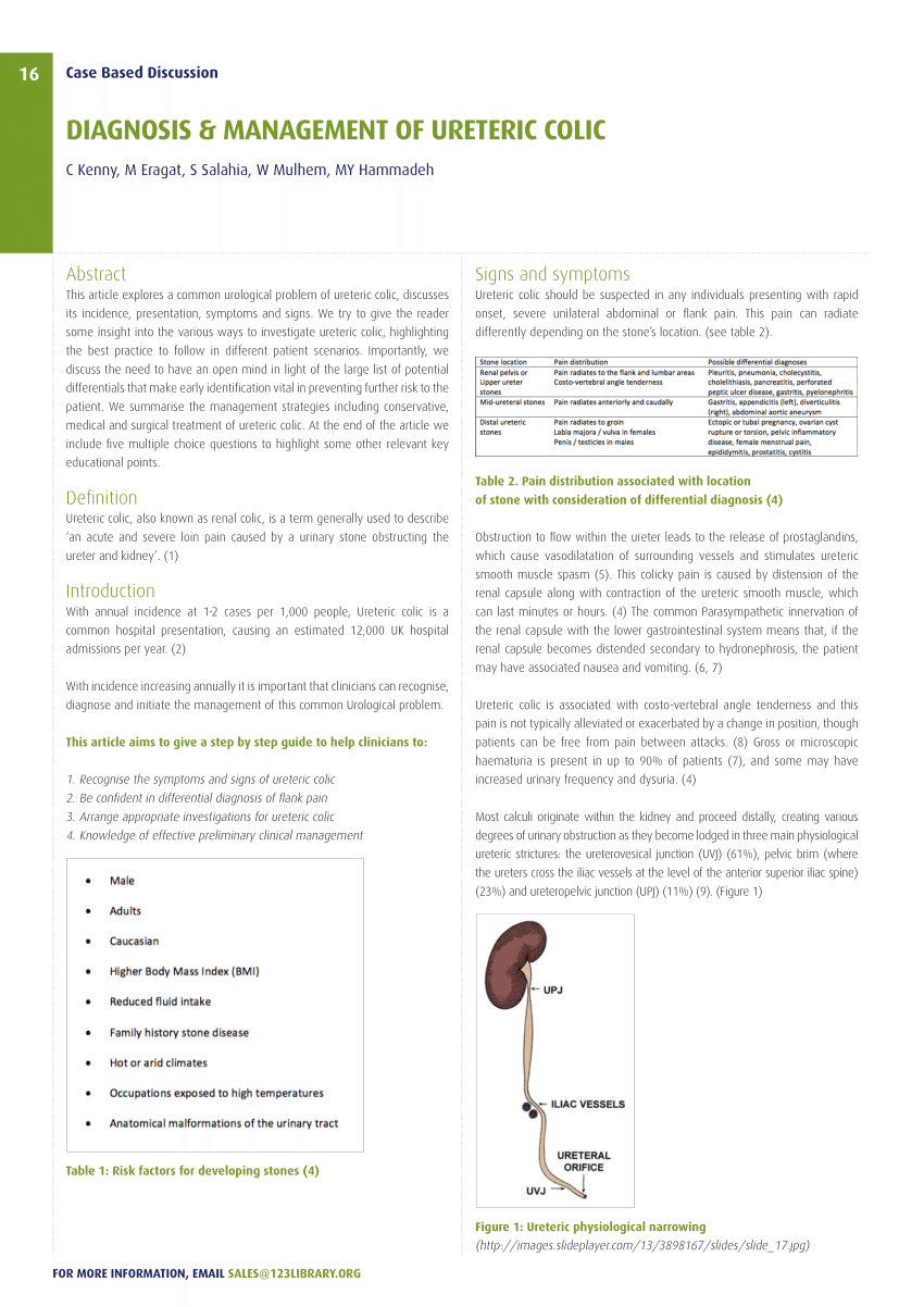
(PDF) Diagnosis & Management Of Renal Colic
Introduction. Renal colic describes intense wave-like pain related to the passage of ureteric stones.It is a very common condition, with an estimated 12% of men and 6% of women experiencing renal colic at least once in their lifetime 1.Most stones are calcium-based (oxalate, phosphate or mixed). Other stones include urate, struvite and cysteine. The following article will cover the assessment.

Renal Colic Urology
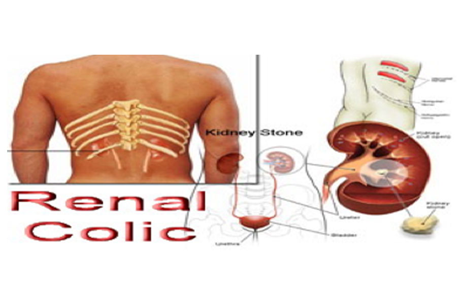
Symptoms of renal colic include: intense pain along the side of your body between your ribs and hip, or in your lower abdomen. pain that spreads to your back or groin. nausea or vomiting. Renal.

Table 2 from Diagnostic management of renal colic. Semantic Scholar
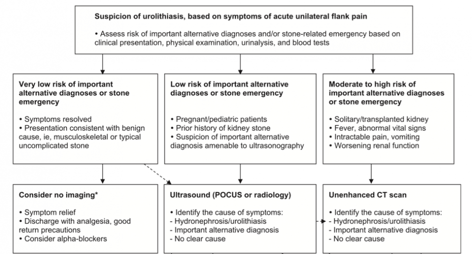
Renal colic is a common reason for presentation to emergency departments, and imaging has become fundamental for the diagnosis and clinical management of this condition. Ultrasonography and particularly noncontrast computed tomography have good diagnostic performance in diagnosing renal colic. Radiologic management will depend on the tools.

Management of Renal Colic and Triage in the Emergency Department Abdominal Key
Calculus of kidney with calculus of ureter. N20.2 is a billable/specific ICD-10-CM code that can be used to indicate a diagnosis for reimbursement purposes. The 2024 edition of ICD-10-CM N20.2 became effective on October 1, 2023. This is the American ICD-10-CM version of N20.2 - other international versions of ICD-10 N20.2 may differ.
Renal Colic
Unspecified renal colic N23-. Clinical Information. A disorder characterized by paroxysmal and severe flank marked discomfort radiating to the inguinal area. Often, the cause is the passage of kidney stones. A severe pain in the lower back radiating to the groin, scrotum, and labia which is most commonly caused by a kidney stone (renal calculus.
Renal Radiology Key
Browse all the diagnosis codes used for unspecified renal colic (n23). For easy navigation, the diagnosis codes are sorted in alphabetical order and grouped by sections. Each section is clearly marked with its description, and the corresponding three-digit code range.. Renal Colic - A severe intermittent and spasmodic pain in the lower back.

Renal colic fast track pathway. Abbreviations AAA, abdominal aortic... Download Scientific
Urolithiasis. ( N20-N23) Calculus of kidney and ureter. ( N20) N20.0 is a billable diagnosis code used to specify a medical diagnosis of calculus of kidney. The code is valid during the current fiscal year for the submission of HIPAA-covered transactions from October 01, 2023 through September 30, 2024.
Renal Calculi Icd 10 ManuelqoHogan
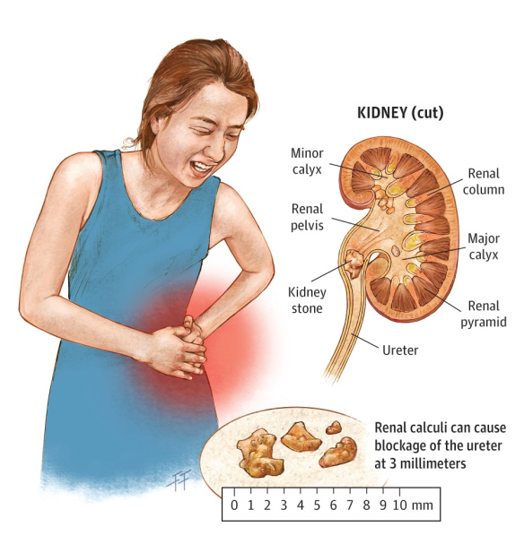
Renal colic is the acute onset of severe abdominal / flank pain due to kidney stones. Pain typically radiates from the flanks to the groin and intensifies over the first 30-60 minutes. Eliciting a history of prior nephrolithiasis, a family history of kidney stones, or patients at risk of nephrolithiasis (eg, gastric bypass surgery, short bowel.

Renal Colic Kidney Stone pain Iqbal Health Centre YouTube
R10.83 is a billable/specific ICD-10-CM code that can be used to indicate a diagnosis for reimbursement purposes. The 2024 edition of ICD-10-CM R10.83 became effective on October 1, 2023. This is the American ICD-10-CM version of R10.83 - other international versions of ICD-10 R10.83 may differ. ICD-10-CM Coding Rules.
Diagnosis and Management of Acute Renal Colic An Evidence Based Update EMOttawa Blog
Unspecified renal colic. N23 is a billable/specific ICD-10-CM code that can be used to indicate a diagnosis for reimbursement purposes. The 2024 edition of ICD-10-CM N23 became effective on October 1, 2023. This is the American ICD-10-CM version of N23 - other international versions of ICD-10 N23 may differ.

CME 30/10/14 Renal Colic Investigation and Management Charlie's ED
This pain is often known as renal colic and typically comes in waves lasting 20 to 60 minutes. Other associated symptoms include: nausea, vomiting, fever, blood in the urine, pus in the urine, and painful urination.. ICD 9 Codes: 592.0, 592.1, 592.9: A kidney stone, 8 millimeters (0.3 in) in diameter .
A Nutshell Review On The Therapeutics Of Renal Calculi
500 results found. Showing 1-25: ICD-10-CM Diagnosis Code N23 [convert to ICD-9-CM] Unspecified renal colic. Renal colic; Renal colic (pain from kidney stone); Ureteral colic; Ureteric colic. ICD-10-CM Diagnosis Code R10.83 [convert to ICD-9-CM]

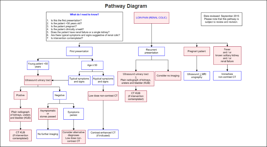
Managing acute renal colic across the primarysecondary care interface a pathway of care based
Objectives Symptomatic ureterolithiasis (renal colic) is a common Emergency Department (ED) complaint. Variation in practice surrounding the diagnosis and management of suspected renal colic could have substantial implications for both quality and cost of care as well as patient radiation burden. Previous literature has suggested that CT scanning has increased with no improvements in outcome.